Services >
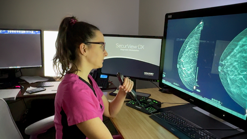
3D mammography is an advanced form of breast imaging that captures multiple low-dose images of the breast from different angles to create a three-dimensional view. This technology allows radiologists to examine breast tissue one layer at a time, improving the detection of breast cancer while reducing the chance of being called back for additional imaging. This technology is used for both routine screening and diagnostic evaluations.
A DEXA scan measures bone mineral density to assess the risk of osteoporosis and fractures. The exam is quick, painless, and uses a very low dose of radiation. Early detection of bone loss allows your physician to recommend treatments and lifestyle changes to help protect your bone health.
A breast biopsy is a minimally invasive procedure used to collect a small sample of breast tissue for laboratory analysis. Most breast biopsies are performed using imaging guidance, allowing the radiologist to precisely target the area of concern while minimizing discomfort and avoiding surgery in many cases.
Breast MRI uses a powerful magnet and radio waves to create detailed images of the breast without using ionizing radiation. It is often recommended for women at high risk for breast cancer, to evaluate the extent of a known cancer, or to investigate findings from other breast imaging exams. Breast MRI provides highly detailed information that complements mammography and ultrasound.
Breast ultrasound uses sound waves to evaluate breast tissue and further assess areas of concern identified during a physical exam or mammogram. It is commonly used to distinguish between fluid-filled cysts and solid masses and to evaluate breast symptoms. Ultrasound also helps guide certain breast biopsy procedures.
A diagnostic mammogram is performed to evaluate breast symptoms, such as a lump, pain, nipple discharge, or an abnormal finding on a screening mammogram. Additional images are taken to provide a more detailed evaluation of the area of concern. The results help determine whether further testing or treatment is needed.
An HSG is a specialized fluoroscopic exam that uses contrast material and X-rays to evaluate the uterus and fallopian tubes. It is commonly performed as part of an infertility evaluation to determine whether the fallopian tubes are open and whether the uterine cavity appears normal. The exam provides valuable information to help guide fertility treatment and other gynecologic care.
A hysterosonogram is a specialized pelvic ultrasound performed after a small amount of sterile saline is placed into the uterus. The saline gently expands the uterine cavity, allowing the physician to better evaluate the uterine lining for polyps, fibroids, scar tissue, or other abnormalities. This exam is commonly used to investigate abnormal uterine bleeding and infertility.
An MRI-guided breast biopsy uses MRI imaging to locate breast abnormalities that cannot be seen with mammography or ultrasound. A small tissue sample is collected for laboratory evaluation using a minimally invasive technique. The procedure helps provide an accurate diagnosis while avoiding surgery in many cases.
An obstetric ultrasound uses sound waves to monitor the health and development of a pregnancy. It provides detailed images of the fetus, placenta, and uterus throughout each stage of pregnancy. The exam helps assess fetal growth, anatomy, and overall well-being while supporting your healthcare provider in prenatal care.
A pelvic ultrasound uses sound waves to examine the uterus, ovaries, cervix, and other pelvic structures. It helps evaluate pelvic pain, abnormal bleeding, ovarian cysts, fibroids, and other gynecologic conditions. Depending on the reason for the exam, images may be obtained through the abdomen, transvaginally, or both.
A screening mammogram is a routine exam used to detect breast cancer before symptoms develop. Regular mammograms can identify breast changes at an early stage, when treatment is often most effective. Annual screening is recommended for most women beginning at age 40, unless otherwise advised by their healthcare provider.
A stereotactic breast biopsy uses mammography images to precisely locate and sample suspicious breast tissue. It is commonly performed to evaluate abnormalities such as calcifications or areas seen only on a mammogram. This minimally invasive procedure often eliminates the need for surgical biopsy.
An ultrasound-guided breast biopsy uses real-time ultrasound imaging to guide a needle into a breast abnormality. The procedure is minimally invasive and allows tissue samples to be obtained with precision. The samples are then analyzed to help determine whether the area is benign or cancerous.